Showing posts with label ER nursing ICU Nursing. Show all posts

Tuesday, June 4, 2024

Management of Sepsis


Management of Sepsis


Sepsis is a critical medical condition caused by organ dysfunction due to an abnormal response to infection. 

The Goals of treatment are: 

 To reduce mortality in adult and pediatric patients with severe sepsis and septic shock by developing a protocol-based approach to case identification and rapid treatment. 

Rapid identification and communication regarding patients with sepsis, severe sepsis and septic shock. 

Treatment measures include: • Blood culture before antibiotics • Antibiotics • Lactate level • Fluid resuscitation • Use of vasopressors if persistent hypotension


Sepsis Definitions


Sepsis Bundles


Effectively managing sepsis and septic shock involves a team effort from healthcare professionals including doctors, nurses, patient care technicians, and pharmacists, among others, to improve patient-centered care and outcomes. 
The most effective way to reduce in-hospital mortality in sepsis patients is through early identification and prompt implementation of treatment bundles according to your hospital's protocols.

Sources: 

Severe Sepsis and Septic Shock: Management Bundle Measure (cms.gov)

Pearls and Pitfalls: CMS and Sepsis – CriticalCareNow

Monday, June 3, 2024

Did You Know That Bruce Lee Died of Heat Stroke? Let's Learn How to Manage Heat Related Illnesses

 How to Manage Heat Related Illnesses


Did you know that martial arts movie icon Bruce Lee died of a heat stroke at the age of 32 on July 20, 1973, the hottest day of the month that year in Hong Kong

Several months before his death, Lee underwent surgery to remove the sweat glands from his armpits, believing that sweaty armpits looked unattractive on screen. This surgery reduced his body's ability to cool itself. On May 10, ten weeks before his death, Lee went into a small dubbing room to re-record dialogue for "Enter the Dragon." 

The sound engineers turned off the air conditioner to prevent noise interference with the recording. After about 30 minutes in the stifling room, Lee fainted and began convulsing. He was rushed to the hospital and nearly died from cerebral edema, which the doctors managed to diagnose and treat just in time.


The medical team did not recognize that his collapse was most likely caused by heat stroke, a common cause of death among young athletic men during the summer. In the United States, an average of three high school and college football players die each year from heat stroke. Autopsies of heat stroke victims often reveal cerebral edema. 

According to Dr. Lisa Leon, an expert in hyperthermia at the U.S. Army Research Institute of Environmental Medicine, “A person who has suffered one heat stroke is at increased risk for another. Patients experience multi-organ dysfunction during the hours, days, and weeks of recovery, which increases the risk of long-term disability and death.”


Heat Related Illnesses

Heat-related illnesses vary in severity from mild to life-threatening. Additionally, heat exposure worsens many common health conditions such as heart, respiratory, and kidney diseases. As summer is drawing near, we have to be aware of heat related illnesses and its management.


Heat Syncope: This refers to dizziness or fainting due to standing up too quickly in the heat or pooling of blood in the limbs from vasodilation, especially when no other cause is identified. It is more frequent in individuals with heart conditions or those taking diuretics. Treatment: Remove the patient from the heat and treat with rest in a supine position, passive cooling, and rehydration (either oral or intravenous). If recovery is prolonged or if there are concerns about a cardiac cause, especially in patients with cardiac risk factors, further evaluation is necessary.


Heat Edema: This is a mild swelling in dependent areas of the body, typically seen in people who are not yet acclimated to hot environments.  Treatment: Remove patient from heat and elevate the legs. Diuretic agents are not indicated.


Heat Cramps: Painful muscle spasms in the abdomen, arms, or legs can occur during or after activity in the heat, often due to excessive salt loss from sweating during physical exertion. Treatment: Remove patient from heat, treat with rest, oral electrolytes, and fluid repletion.


Heat Exhaustion (Prostration) - Profound fatigue, weakness, nausea, headache, or dizziness (or a combination of these symptoms) can result from decreased body water content or blood volume due to water or salt depletion from heat exposure. Mild body temperature elevation (below 40°C) may be present, but there is no altered mental status. Treatment: Remove the patient from the heat and treat with rest in a supine position, evaporative cooling, and rehydration (intravenous or oral). Monitor mental status, and if there is a delayed response to treatment, further evaluation is needed.


Heat Stroke

Heat stroke is the least common but most dangerous heat illness, often fatal without immediate treatment. The most frequent victims are the urban elderly poor. It is identified by a body temperature exceeding 40ºC.

    Predisposing Factors: Salt and water depletion, infections, fever post-immunization, lack of acclimatization, obesity, drug use, fatigue, and various medical conditions like cardiovascular disease, diabetes, malnutrition, alcoholism, hyperthyroidism, impaired sweating, and potassium deficiency. 

Heat stroke can be categorized into two forms: classic and exertional.

  1. Classic Heat Stroke: Occurs after several days of high temperatures (above 37.0ºC) or lower temperatures with high humidity. Continuous sweating eventually ceases, causing a rapid rise in body temperature. The first symptom may be sudden collapse. In 80% of cases, onset is abrupt with the patient becoming delirious, comatose, or having seizures. In the remaining 20%, symptoms like weakness, dizziness, nausea, fainting, or frontal headache precede collapse. Muscle cramps are rare. Patients typically have hot, dry skin but may continue sweating. Progression to coma is common.

  2. Exertional Heat Stroke: Occurs with activities that increase internal heat production. In the U.S., it commonly affects long-distance runners, football players, and military recruits. Intense physical activity usually precedes heat stroke. Overuse of salt supplements with insufficient water can be fatal. Core temperatures might not exceed 40ºC and still be lethal, while in classic cases, patients have survived with temperatures as high as 43.7ºC. Rapid cooling is crucial to prevent extensive tissue necrosis. Common complications include thrombocytopenia, prolonged PT and PTT, marked DIC, hypotension, shock, acute pulmonary edema, and rhabdomyolysis. Severe hypoglycemia and death often result from acute hyperkalemia or renal failure due to extensive muscle breakdown.

Heat Stroke Management: Immediate cooling is critical. Delays in cooling due to misdiagnosis, underestimation of severity, or lack of facilities are major factors leading to death or disability. The ABCs (Airway, Breathing, Circulation) are the first priority. Effective cooling methods include ice water immersion or evaporative cooling with fans and skin wetting. Cooling should cease when core temperature reaches 39º-40ºC, as body temperature will continue to fall. Avoid large initial IV fluid amounts due to the risk of pulmonary edema; use fluid boluses with careful CVP and urine output monitoring. Avoid alpha-adrenergic drugs like norepinephrine. Check glucose levels and administer D50% if needed, especially in exertional heat stroke. Severe rhabdomyolysis may cause hyperkalemia and myoglobinuria, leading to renal failure, shock, and diaphragm necrosis with respiratory failure. Hyperthermia may recur within 3-4 hours, requiring repeated cooling. ICU admission is warranted for management of end-organ sequelae. Monitor patients closely as they may lose the ability to sweat for several weeks.


References:

Friday, May 31, 2024

Adult Lumbar Puncture (LP) Procedure for Diagnostic Testing


Adult Lumbar Puncture (LP) Procedure for Diagnostic Testing 


Indications 

For diagnosis of:  Subarachnoid hemorrhage, Meningitis, Neurological disorders, Cerebral lymphoma / metastatic disease 


Contra indications 

Bleeding disorders (e.g. PT > 1.3, Platelet count < 80,000), recent heparin, LMWH or other anticoagulant 

 Local sepsis such as cellulitis or abscess on back 

 Raised intracranial pressure 

 Note indications for CT scan prior to LP above 

 Previous back surgery (relative) 

 Prior history of back pain / pathology (relative) 


In certain conditions, a brain CT has to be done first before an LP is performed: 

 Immunocompromised patients 

  Recent history of head trauma 

  Impaired level of consciousness 

  Evidence of papilledema or focal neurological deficits

 If a lumbar puncture (LP) is delayed to conduct neuroimaging studies, empirical antibiotic therapy should begin after obtaining blood cultures. It is crucial to perform the LP, if there are no contraindications, and start antibiotics as soon as possible in a patient with suspected meningitis, as the outcome is dependent on the timeliness of the treatment initiation.


Supplies needed

 LP kit 

 Includes 23 g 9cm spinal needle 

 Mask, gown, gloves 

 Assistants- to hold the patient in position and to hand equipment to the operator


Consent 

Patient consent must be secured, and the procedure should be thoroughly explained to them. Major complications can involve post-dural puncture headaches, while significant back pain is rare.


 Operator (the person doing the LP)

Follow your hospital's GME policy for residents performing LP. In some city hospitals a resident has to complete 3 to 5 successful LPs with an attending doctor present. 


Patient position 

For right-handed practitioners, patients should be positioned on their left side, though a sitting position is also an option. In the sitting position, patients should sit at the edge of the bed or trolley with their legs hanging over the side. An assistant can help support the patient. Placing a pillow on the patient's lap can be beneficial, with the patient leaning forward over it.


Lumbar Puncture Procedure

The operator should wear a surgical mask, gown, and gloves after performing a surgical scrub. The patient's back should be prepared with Betadine or a similar antiseptic and allowed to dry.

Once the skin preparation is dry, the area should be covered using the plastic drape that comes with the kit, or alternatively, sterile sheets can be used to demarcate the prepared area.

The L3/4 interspace is identified using the intercristal line as a surface marker (the line at the level of the iliac crests). The spinal cord typically ends at the lower border of L1; lumbar punctures should be performed below this level.

A small amount of local anesthetic is injected into the skin and subcutaneous tissues. A 23-gauge spinal needle with a stylet is then inserted perpendicular to all planes at the L3/4 interspace and advanced into the subarachnoid space, which can be felt as a slight pop. 

The depth to the space is approximately 5 cm but can range from 3 cm to 9 cm in larger patients. If bone is encountered, withdraw the needle almost to the skin and try another approach, either more cephalad or caudad. Do not force the needle, as it can bend or fracture. Replace the stylet with each advancement of the needle. If there are three unsuccessful attempts, the operator should seek assistance. 

Remove the stylet and check for CSF flow. If CSF is present, use the manometer to measure opening pressure, considering the patient's position during interpretation. I possible, opening pressure should be measured. It is usually elevated in cases of bacterial or cryptococcal meningitis. 


Collect the initial CSF that emerges into the first tube and continue to collect CSF into pre-numbered tubes in the correct order. After collection, remove the needle and apply an appropriate dressing. Many emergency department texts recommend replacing the stylet before removing the needle. Ensure the needle is complete and intact after removal.


Cerebrospinal Fluid Collection Tubes

Tube one, which is potentially the most contaminated and blood-stained, is sent to biochemistry. Tube two is sent to cytology, and tube three, the least likely to be contaminated or blood-stained, is sent to microbiology. By keeping this in mind, it may be possible to collect additional CSF in certain tubes based on the clinical presentation. For example, in an adult with suspected meningitis:


 Always collect CSF into the tubes in the correct order (i.e. tube 1 first; tube 3 last). Some facilities require a 4rth tube to be collected which is used for comparison if result of any of the 3 other tubes need to be confirmed rather than sticking the patient again. 


(Note: Always follow your facility's policy and procedure on lumbar puncture for diagnostic tests.)


Monday, May 27, 2024

Administering Tissue Plasminogen Activator (TPA) for Ischemic Stroke

 ADMINISTERING TISSUE PLASMINOGEN ACTIVATOR (TPA) FOR ISCHEMIC STROKE


Stroke is a clinical syndrome, believed to be of vascular origin, presents with sudden signs of localized or widespread brain dysfunction lasting more than 24 hours and may result in disability or in death. This can manifest as ischemic cerebral infarction (about 87% of cases) or as intracerebral hemorrhage (about 13%).

One of the treatments for ISCHEMIC STROKE is thrombolytic therapy. 

Candidates for thrombolytic therapy are given the appropriate therapy within the shortest time possible based on evidence-based guidelines such as the AHA. 

The window of opportunity for eligible patients to receive thrombolytic therapy for acute ischemic stroke is THREE to FOUR AND A HALF hours from a clearly defined time last known well or the onset of signs/symptoms

The door-to-needle time (time of bolus administration) should be within 60 minutes from hospital arrival.

IV rTPA Administration: 

0.9 mg/kg to a maximum of 90 mg

  First 10% of calculated dose given as intravenous bolus dose 

 Remaining 90% of calculated dose given in infusion over 1 hour 

o Verify that the Physician has reviewed the inclusion/exclusion criteria and discussed the plan with the patient and/or family if available. 

o Verify that administration will start within three hours of symptom onset or time last known well (Verbal Consent of pt./family). If administering after 3 hours, a written consent is needed. Follow your hospital's policy.


Equipment Needed:

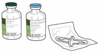
o 1 vial of Alteplase (Activase) 50mg or 100 mg 

o One 10 ml syringe

o 50ml syringe and vented spike connector for the 50mg vial (does not come with kit)

o Two 19-gauge needles

o One blunt cannula

o Standard pump tubing

o Intravenous infusion pump

o 100 cc bag of 0.9% NS

o Alcohol wipes

o Two Medication labels


Administration Procedure:

o Verify the bolus dose and the infusion dose.

o Reconstitute the vial of t-PA with the supplied preservative-free water

o Direct stream of water into lypophilized cake

o Swirl but DO NOT SHAKE (slight foaming is common)

o Let stand several minutes to allow large bubbles to dissipate

o Final concentration is 1 mg/ml

o Fill out IV medication label with all required information (patient name, medication, dosage, time, date, RN signature) and affix label to Alteplase bottle. 

o Draw waste dose from bottle and verify waste amount by showing to the Stroke 

Neurologist/Physician and another nurse.

o Connect Activase bottle to IV pump tubing, carefully priming to avoid discarding any medication.

o Verify patency of IV site and tubing connections

o Verify that all blood work has been drawn and sent

o Attach noninvasive blood pressure cuff to other arm

o Set infusion pump rate for bolus dose to be given over 1 minute and infusion dose according to 

dosing sheet with a total infusion time of 1 hour.

o For manual bolus administration (as requested by the Physician/Neurologist), using a 10 ml syringe, withdraw the bolus dose directly from the Activase bottle (see dosing sheet for bolus dose based on patient weight*). 

o Fill out medication label with all required information (patient name, medication, dosage, time, date, RN signature). Hand the bolus dose syringe to the Stroke Neurologist/Physician.

o Ensure that the patient’s systolic is <180 and diastolic is <110 prior to and during infusion.

o Document infusion start time and name of Physician/Stroke Neurologist (as needed).

o When pump alarms "no flow above", there is still some medication left in the tubing which must be infused. Remove the IV tubing connector from the Activase bottle and attach it to a newly spiked 100 cc bag of 0.9% NS.

o Continue the infusion at the current setting to deliver the remainder of the original Activase volume over the remaining time. Continue the infusion until the preset volume is completed.

o Document end time of infusion. 

Monitoring

Once infusion begins monitor vital signs and neurologic status as follows:

 Every 15 min for 2 hours, then:

 Every 30 minutes for 6 hours, then:

 Every 60 minutes for 16 hours

*Notify physician immediately if SBP/DBP greater than 175/100

Precautions and Side Effects

 √Hemorrhage (GI, GU, catheter puncture site, intracranial, retroperitoneal, pericardial, 

gingival, epistaxis). NO blood works for 24 hours (except for emergent lab.).

 √New ischemic stroke

 √ Bruising

 √Anaphylaxis

 √Laryngeal edema

 √Rash, urticaria


o If the patient’s neurologic status declines during tPA infusion the following actions should be taken:

  Stop the infusion 

 Page the Stroke Neurologist 

 Draw and send PT/PTT, D-Dimer and fibrinogen 

 Prepare for emergent CT


NOTE: Always follow your hospital policy on TPA administration.


References: guidelines-for-mangaging-patients-with-ais-2019-update-to-2018-guidelines.pdf (stroke.org)

Acute Ischemic Stroke Resources | American Stroke Association

Saturday, May 25, 2024

How to administer Tranexamic Acid or TXA for Bleeding in Trauma


Tranexamic Acid or TXA for Bleeding in Trauma

Tranexamic Acid or TXA is an anti-fibrinolytic drug, a synthetic equivalent of the amino acid lysine, and acts opposite to tissue plasminogen activator or rTPA. This is why TXA is sometimes called anti-TPA.

TXA can help to reduce mortality in the acute severely bleeding adult trauma patient if given within three hours of injury.  

Contraindications for its use are acquired defective color vision hypersensitivity to TXA or any of its ingredients. Do not give in conjunction with prothrombin complex concentrate such as K Centra, active intravascular clotting and or history of thromboembolism and renal failure.

Indications for use of TXA in the bleeding adult trauma patient are: Administration less than three hours from time of injury, severe hemorrhagic shock with systolic blood pressure below 90 millimeters mercury, heart rate above 110 beats per minute, multi-system trauma with evidence of active hemorrhage, major pelvic fracture with evidence of active hemorrhage, solid organ injuries with evidence of active hemorrhage, and traumatic amputation

How to administer TXA. 

A loading dose of one gram TXA is reconstituted in 100 milliliters of normal saline and given intravenously over 30 minutes. 

Followed by an intravenous infusion of one gram TXA mixed in 250 ml normal saline over 8 hours. 

Watch for adverse side effects such as anaphylaxis, thrombosis, nausea, vomiting, diarrhea, visual disturbances, and hypotension. Stop the infusion and notify the doctor immediately. Follow hospital protocol. 

Click the link below for a video about TXA. Thank you.

https://www.youtube.com/watch?v=Mzxn1gA82j4 

Sickle Cell Disease (SCD)

    Sickle Cell Disease (SCD) Sickle cell disease (SCD) is a collection of genetic blood disorders in which abnormal levels of hemoglobin, c...